During our summer vacation of 2012, our pet-sitter called us to say that there was a problem with Dashy. They went on explained that he was not very playful during the previous days, but they could not find anything physically wrong with him. That day, after spending hours hidden, they realized that he had a white, tick membrane on his eye. They promptly took him to the vet and called us to inform that Dashy would need a medical intervention.
We arrived in the next day, took him to his regular vet who advised us to take him to a specialist in ophthalmology. The ophthalmologist examined Dashy and explained to us that he had a disease called distichiasis: an abnormal growth of eyelashes at the eyelid margin, which grow in the direction of the eye. This is a common disease in Shih Tzus and other breeds like Cavalier King Charles Spaniel, Lhasa apso, or Bulldog (to cite a few). Because we could not identify the problem in its early stages, the corneal irritation turned into a deep ulcer.

During the examination, the vet also discovered that both eyes had distichiasis, but only the left eye had developed an ulcer. If not treated correctly, corneal ulcers can lead to blindness and even loss of the eye. The picture on the left show the distichiasis on Dashy's left eye (no ulcer formed). With the eyelashes irritating the eye, an abundant discharge is produced (lower right corner of the eye).
The suggested treatment was to perform surgery in order to:
- remove the problematic eyelashes and kill the hair follicles to avoid the distichiasis to return;
- place of a conjunctival pedicle graft into the corneal ulcer. A very good description of such procedure can be found in Willow's Veterinary Centre & Referral Service. According to their website:
The conjunctiva is a thin membrane that covers the white of the eye. The conjunctiva has many blood vessels in it, although in the normal eye it is only lightly pink. [...] In cases of rapidly progressive corneal ulcers, it is possible to surgically create a stalk or ‘pedicle’ of conjunctiva which can then be stitched into the corneal ulcer. The pedicle of conjunctiva not only provides some stability for the weakened cornea but also brings blood vessels (carrying healing cells and antibodies) right to the ulcer – this helps to fight infection.
During surgery, the vet removed the problematic eyelashes, but also found out that Dashy had, as well, ectopia cilia. Ectopia cilia is a disease in which single or multiple hairs grow through the inside of the eyelid. Distichiasis and Ectopia cilia could be found in both of Dashy's eyes, and were removed by the vet.
.JPG) If you are interested in seeing the pictures of the corneal ulcer and the conjunctival pedicle graft, as well as the evolution of the treatment, please click here. Warning: the images are not very pleasant to see.
If you are interested in seeing the pictures of the corneal ulcer and the conjunctival pedicle graft, as well as the evolution of the treatment, please click here. Warning: the images are not very pleasant to see.
Dashy then spent several weeks wearing a protective collar. He looked a little bit down after the surgery, but some days later he was already used to the collar and running around.
Battles against Distichiasis and Ectopia cilia
As explained by the vet, killing the hair follicles would avoid the distichiasis to return in that particular place, but would not avoid it to grow in other follicles. Unfortunately, two weeks after the surgery, during an examination, the vet found more distichiasis and ectopia cilia growing in both eyes. A new surgery was then performed to remove and kill these follicles.
Since these problems started, we had been paying attention to any sign of eye discomfort. About 3 weeks after the second surgery, we realized that Dashy was presenting excessive blinking, eye discharges, and his right eye was only half opened. We promptly took him to the vet and found out that the distichiasis and ectopia cilia were back, and an ulcer was starting to form on his right eye. Another surgery was performed, but this time, the vet was able to reconstruct the cornea without the need to place a conjunctival pedicle graft. The photos are here. Once again, the offending eyelashes was removed and hair follicles killed.
A couple of months later, Dashy had recovered and the graft was removed, leaving an almost imperceptible scar.
Since then, distichiasis and ectopia cilia have been a recurrent problem. The vet realized that keep removing the hair follicles would not longer be a good solution because it would end up damaging too much the support structure of Dashy's eye. Besides, the follicles are responsible for producing good quality tears. Killing them would affect the lubrication of the eye and aggravate the problem. The solution has been to use an ophthalmic lubricant to protect the cornea and coat the lashes. The idea is to keep the cornea always lubricated, and the eyelashes soft enough to slide on the cornea instead of damaging it.
If some eyelashes start damaging the cornea, the vet now removes them by cryosurgery, which removes the hair, but does not kill the hair follicles.
After this experience, we have been able to detect the signs of big problems or normal irritation due to the growth of new eyelashes. Fortunately, we have managed to space the visits to the vet clinic every 2 months, and Dashy's last surgery was about 6 months ago. =)
Corneal Degeneration
In February 2013, the graft was healing as expected, but a white, crystalline stain had appeared on Dashy's eye. The vet diagnosed it as a corneal degeneration, a disease in which lipid or calcium deposits within the corneal stroma, and/or epithelium. It may occur in areas of the cornea that have suffered traumatic incident or chronic disease process.
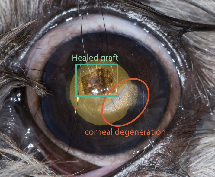
Corneal degeneration can lead to corneal ulcers (superficial or deep), ocular infections, and corneal scarring and vascularization arriving from continuous sloughing of the mineral deposits [1]. The vet decided to observe the evolution of the condition since Dashy was not presenting any discomfort. Unfortunately the degeneration evolved to a vascularization (see pictures here). A topic treatment was prescribed and, in about one month, the vascularization was gone and the mineral deposited dissolved into a less opaque area.
No comments:
Post a Comment